Thursday, November 19, 2015

Forensic Students Investigate and Sketch a Crime Scene

The Forensic Science classes have been examining, documenting and photographing a crime scene in their current unit of study. The students work the scene in teams of two to three students. Each team is given one eighty minute period to document the scene by itemizing, describing and photographing the evidence found in the class room. When completed they will write an investigative report that will include they hypothesis as to what the evidence indicated occurred in the room.

As they move through the scene they will prepare four different crime scene forms:

[1] Crime Scene Disposition Form - in which each piece of physical evidence is assigned a unique identifying number/letter, described and earmarked for specific tests (fingerprints, DNA, drugs etc.) when the evidence is gathered and returned to the lab.
[2] Crime Scene Measurement Form - in which each piece of evidence's position is precisely located by measuring its distance from two points in the room.
[3] Crime Scene Photograph Form - in which each piece of evidence is photographed and that photograph is hyperlinked to the evidence description in the log.
[4] Crime Scene Rough Sketch Form - in which a bird's eye, not to scale replica of the crime scene and all the evidence is produced.
Bloody foot prints leading from the table where an altercation occurred.


Bullet casing.

Left side of table where the homicide victim was found seated.

Right side of table where the homicide victim was found seated.

Spent projectile on window sill behind homicide victim.

Robert catalogs the physical evidence on the table, while Daniel is entering his data into the digital forms.

Jack was given the task of measuring the positions of all the physical evidence. Daniel is assigning item numbers to the evidence, writing descriptions and deciding on what the disposition of the evidence should be once it arrives in the lab.

Jack and Robert examine the ledge behind the homicide victim

Sophia and Jack examine the evidence on the table before the homicide victim.

Monday, October 26, 2015

AP Biology Students Examine Rate of Photosynthesis

In their study of photosynthesis, the AP Biology students are carrying out the "leaf disc assay" in which they cut disks out of plant leaves and use a syringe to force the air in the leaves' sponge spongy mesophyll out replacing it with sodium bicarbonate. They then expose the leaves to light and measure the rate of "leaf floatage" as photosynthesis releases oxygen into the spongy mesophyll causing the disks to float. The students count how many disks are floating at 30 second intervals until all the disks are floating or 10 minutes has elapsed. Their baseline experiment involved comparing the rates of photosynthesis in five different type of leaves: spinach, kale, Acuba japonicum, ivy and a red Coleus.

Mallory gets ready with the "fish bowl" heat sink, while Beth uses the hole punch to produce their leaf disks

Lindsay cuts leaf disk from her sample.

Samantha is loading her syringe with the leaf disks.

Ansley is looking for "leaf floation" in her setup.

Neal cuts disks for the next run, while Thomas watches for successful floataion.

Indira pumps the leaf disks in the syringe to get the air out and sodium bicarb into the disks.

......meanwhile Lindsay is still cutting disks......

Meagan smiles while waiting for her disks to float.

Kaitlin is forcing the air out of her disks as Audrey prepares a new leaf for "disking".

Rachael removes the air from this team's disks, while Liv and Allegra cut addition disks, apparently all of this work is under the watchful eyes of their supervisor, Emily

Maddie and Hadley ca not belief how difficult is is to get the leaf disk to discharge their oxygen, uptake the sodium bicarb which is heavier and SINK!!!

Friday, October 23, 2015

Students in Forensic Course Begin Forensic Entomology Investigation

The forensic classes have just begun the examination of the various stages if insect life cycle in to prepare for the determination the TOD (time of death) for two unidentified bodies in the ongoing case study they will be pursuing over  the course of this year.

Not being true forensic entomologist, who already know how to identify the main necrophagous insects that utilize a victim's body, the students began today by examining exemplars of two different species at various points in their life cycle. They were to observe the stages they were given looking for unique characteristics that would allow them to distinguish and identify the insects.

Once they have done this they will be given the insects found on the victims bodies, which had been located in an isolated cabin. They will identify the insects and use them to determine a time of death for the two victims.
The insect exemplars Thomas examined under the dissecting microscope.

Students worked in teams of four, each pair was responsible for identifying and categorizing one species' life cycle exemplars.

Sara examines an exemplar, while her partner logs the information into the evidence disposition log for their team.

Jill has begun to input the data collected so far into a google form for compilation and sharing. Her partner Emme is taking a picture of the exemplar through the dissecting microscope for future reference.

Kathleen is getting ready to examine the next exemplar, while her partner, Helen examine their photo of the examplar they just finished observing and logging.

Lain re-energizes with sip of her iced coffee (shades of Abby Sciuoto from NCIS and her caf-pow), while Gianna slaves away cataloging the current examplar being examined.

Jack is focused on observing the current exemplar as Liam looks on.

Liam is using the coarse adjustment to bring this exemplar into focus, as Jack looks on.

Lily and Maggie converse about Lily's uncanny ability to take awesome pictures of their exemplars. Lily looks so pleased with her success!!!

Thomas closely examines the maggots in the first picture of this series.

Saturday, October 10, 2015

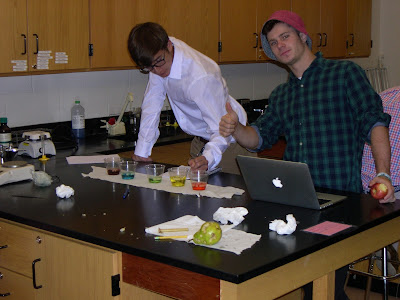
AP Biology students investigate Osmosis, Diffusion and Water Potential

In their study of membranes, osmosis, diffusion and water potential the AP Biology class carried out a number of different investigations. In this extension.open ended investigations, the various teams of students chose to investigate how solutions of differing molarity affected different plant cells (in the guise of different fruits and vegetables. In the baseline protocol the students used potato cores to determine the molarity of each of the different colored solutions. By monitoring the change of mass in the potato cores they were able to accomplish that identification and determine the water potential of the potato cells in the cores.
Jeanne, Freddie, Audrey, Christina and Kaitlin form an awesome team, even if they forget to return the stock solutions in the gallon containers back to the equipment/supply table.

Samuel gives Michael a thumbs up for a job well done.

John watches suspiciously as Maddie pour the blue solution over their cores. Jason must be engrossed in reviewing the protocol they are following.

Rachael and Emily are all smiles, while Liv satisfies her hunger pangs by consuming an extra apple.

Monday, October 5, 2015

Forensic Class Identifies "Skeletal Remains"

In the forensic anthropology unit the students examine and measure the "skeletal remains" of 5 different victims. Most of the samples include a skull, pelvic girdle and at least one long bone (femur, tibia, humerus). Through a thorough examination/measurement of the characteristics of these bone samples, the students should be able to deduce the age, height, sex and ancestry of the individual. The students are also exhibiting their school spirit for the coming weekend's Homecoming Celebration. Go Broncos!!!
Lain is examining a skull. The skull will hold clues to the sex, age and ancestry of this individual.

Emma is entering her skull measurements into a google form to share her data with the rest of the Forensics students.

Liam is proceeding with the measurements of the pubic bone of the pelvic girdle of his team's remains.

Ian and Thomas consult on their observations of the femur from their skeletal remains.
Carlie was so intent on measuring her long bone she was not even aware she was being photographed hard at work.
Martin and Eric are busy using their measurement to calculate the height range of their skeletal remains.
Julia examines the proximal head of the tibia from her skeletal remains, explaining how she will use this information to her teammates. Michelle is intent on reviewing her data tables to assure the are a correct reflection of her observations so far.
Yusrah holds the femur of her team's skeletal remains while Ralph measures its length using the large calipers.
Julia measures the proximal head of the tibia from her skeletal remains.
Cindy uses the large calipers to measure the length of the humerus of the skeletal remains being studied by her team.

Emme and Helen ponder pelvic parameters of their skeletal remains.
Lily and Maggie are examining a pelvic girdle to determine the sex of this individual.


Monday, March 2, 2015

Core Chem Students Learn to Manipulate Bunsen Burner Flame

An important skill for chemistry courses is learning how to control the flame of a Bunsen Burner. Students must maximize the oxygen available to the flame, so that the gas is burned cleanly and provides maximum heat. In this lab exercise, the students learn how to produce such a flame and control its height and positioning. They will use these skills in future lab exercises.


Kathryn positions her burner under the flask to be heated.


Sarah uses her IPad to record the oxygen deficient yellow flame.


Tyler adjusts the main gas valve as Alex looks on.